![PDF] Plant stellate trichomes: strange contaminants appearing in](https://d3i71xaburhd42.cloudfront.net/3ade93a31a0154db40b1e17fcee92bb68361caf3/1-Figure1-1.png)
PDF] Plant stellate trichomes: strange contaminants appearing in
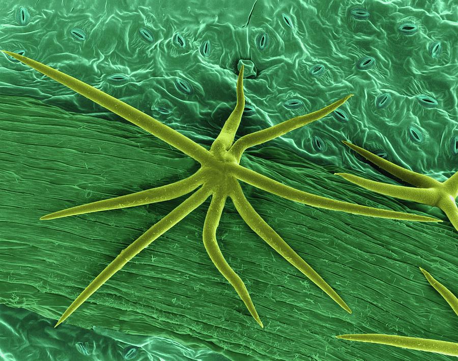
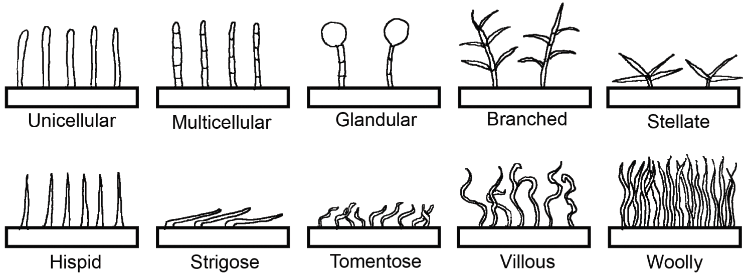
“floral-shaped fibers”, similar in morphology to the stellate structures observed, were reported in skin samples from patients with Morgellons disease and hypothesized by the authors to be keratin fibers produced by keratinocytes. Cornelius and Shelley reported similar findings in 1968. Following careful examination of their possible biological origins, they concluded that these were fragments of plant trichomes, also known as stellate hairs, the unior multicellular outgrowths present on the epidermal surfaces of plant vegetative and floral organs. Based on their morphological characteristics, Cornelius and Shelly proposed that the origin of the stellate trichomes in their preparations were from the undersurface of leaves of Viburnum dentatum, or southern arrowwood. Similarly, Fleischer et al. (1994) identified arrowwood trichomes as nonpathogenic artifacts on skin preparations. Interestingly, these structures have also been observed as contaminants in pap smears. Given the long, tubular nature of the stellate trichome arms these structures may be mistaken for fungal hyphae or other fibrous material. For example, “floral-shaped fibers”, similar in morphology to the stellate structures we observed, were reported in skin samples from patients with Morgellons disease and hypothesized by the authors to be keratin fibers produced by keratinocytes.
Leaf Biophysics (Chapter 2) - Leaf Optical Properties
Scanning electron microscope (SEM) images of the foliar trichomes of

Leaf Paano ito?

The Role of Trichomes in Plant Defense The Quarterly Review of Biology: Vol 48, No 1, Part 1
View PDF - Australasian Plant Pathology Society
Role of Trichomes in Plant Stress Biology

Role of Trichomes in Plant Stress Biology

PDF) Plant stellate trichomes: strange contaminants appearing in KOH preparations

American Journal of Botany
Plant Surfaces: Structures and Functions for Biomimetic Innovations

Leaf - Wikipedia

PDF] Plant Fiber Contaminants in Morgellons Disease Skin Samples