Comparison of the morphologies of adult and embryonic shells of Sepia
Download scientific diagram | Comparison of the morphologies of adult and embryonic shells of Sepia officinalis. a, b Views of an adult shell showing the main morphological parts, a ventral view, b dorsal view. c Dorsal view of a stage 29 embryonic shell. d–f In situ localisation of embryonic shells, d Lateral view on a stage 27 embryo (X-ray image, note the mineralized statocysts anteriorly—st), e dorsal view on a stage 27 embryo, f dorsal view on a stage 29 embryo. All the shells are shown in the same position, at the top: anterior part, on the bottom: posterior part (a–c photographs, e, f optical images, d X-ray image) from publication: Comparison of embryonic and adult shells of Sepia officinalis (Cephalopoda, Mollusca) | Development and evolution of the shell in cephalopods is difficult to establish as there is few species with a calcified shell that could be fossilized (stable in geological time). Internal cuttlebone of sepiids is so particular that homologies are difficult to find. The | Shell, Cephalopoda and Mollusca | ResearchGate, the professional network for scientists.

Comparison of the morphologies of adult and embryonic shells of Sepia

A dEvELopMENTAL TABLE oF EMBryogENEsIs IN Sepia officinaliS

Biology, Free Full-Text

4680 PDFs Review articles in CEPHALOPODA

Organogenesis of embryonic development of Sepia pharaonis illustrating

Comparative brain structure and the neural network features of cuttlefish and squid

Yannicke DAUPHIN, collaborator, PhD and Habilitation
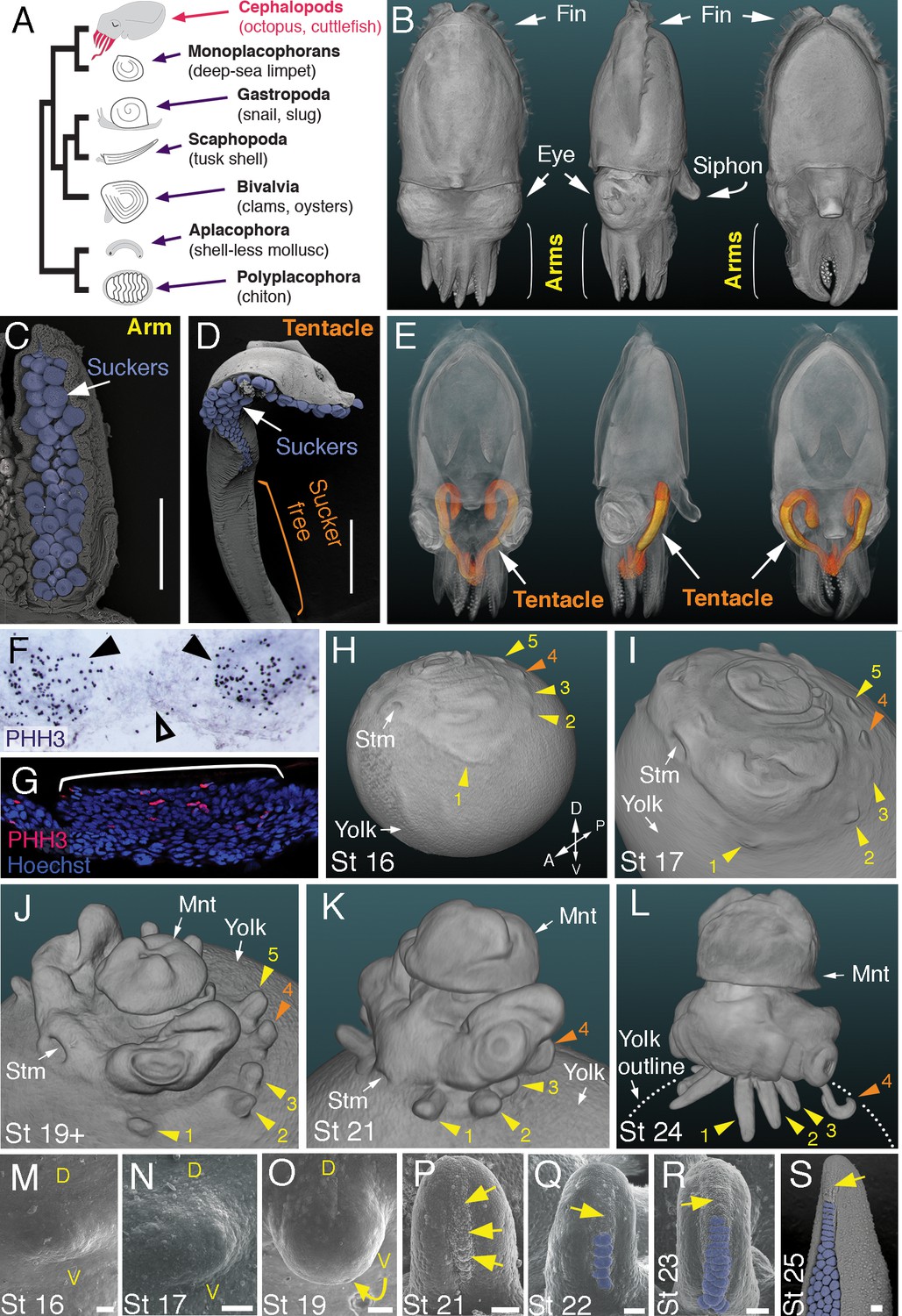
Figures and data in Evolution of limb development in cephalopod mollusks
Biology, Free Full-Text

10 Micro-ornamentation on the embryonic shell of the Ammonitina.