Comparison of the morphologies of adult and embryonic shells of
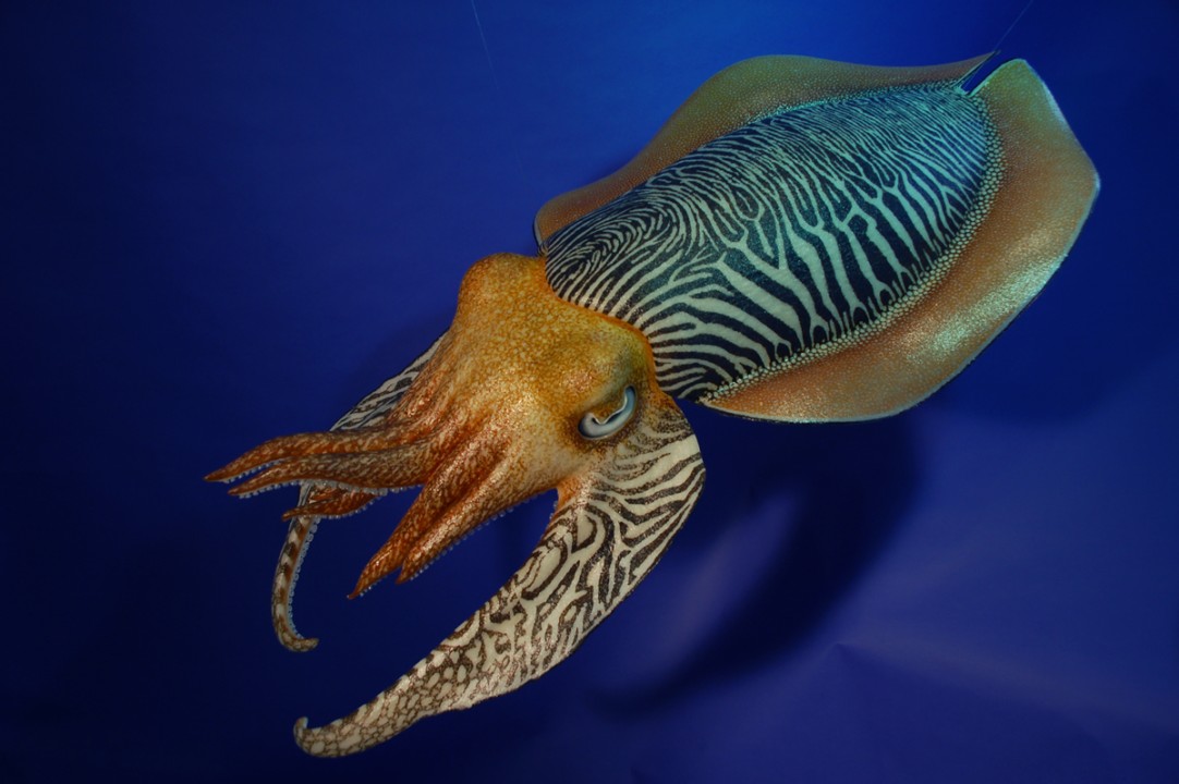
Download scientific diagram | Comparison of the morphologies of adult and embryonic shells of Sepia officinalis. a, b Views of an adult shell showing the main morphological parts, a ventral view, b dorsal view. c Dorsal view of a stage 29 embryonic shell. d–f In situ localisation of embryonic shells, d Lateral view on a stage 27 embryo (X-ray image, note the mineralized statocysts anteriorly—st), e dorsal view on a stage 27 embryo, f dorsal view on a stage 29 embryo. All the shells are shown in the same position, at the top: anterior part, on the bottom: posterior part (a–c photographs, e, f optical images, d X-ray image) from publication: Comparison of embryonic and adult shells of Sepia officinalis (Cephalopoda, Mollusca) | Development and evolution of the shell in cephalopods is difficult to establish as there is few species with a calcified shell that could be fossilized (stable in geological time). Internal cuttlebone of sepiids is so particular that homologies are difficult to find. The | Shell, Cephalopoda and Mollusca | ResearchGate, the professional network for scientists.

Cells, Free Full-Text

DOC) Parasite: SCHISTOSOMA Leah Murray
Comparison of the morphologies of adult and embryonic shells of Sepia

Cells, Free Full-Text

Cells, Free Full-Text

Comparison of the morphologies of adult and embryonic shells of Sepia

Laure BONNAUD, Professor (Full), Muséum National d'Histoire Naturelle, Paris, Adaptations du Vivant- UMR BOREA MNHN/CNRS7208/SU/IRD207/UCN/UA

Embryonic and adult shell groups of Tylomelania. a-c

Comparison of embryonic and adult shells of Sepia officinalis (Cephalopoda, Mollusca)

A; pre-vitellogenic atretic oocyte, pre-vitellogenic normal oocyte and

Comparison of R1 and Atoh1/ nGFP mESC Developmental Morphologies

Comparison Of Embryonic And Adult Shells Of Sepia, 49% OFF

Human embryonic development: from peri-implantation to gastrulation: Trends in Cell Biology