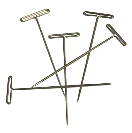
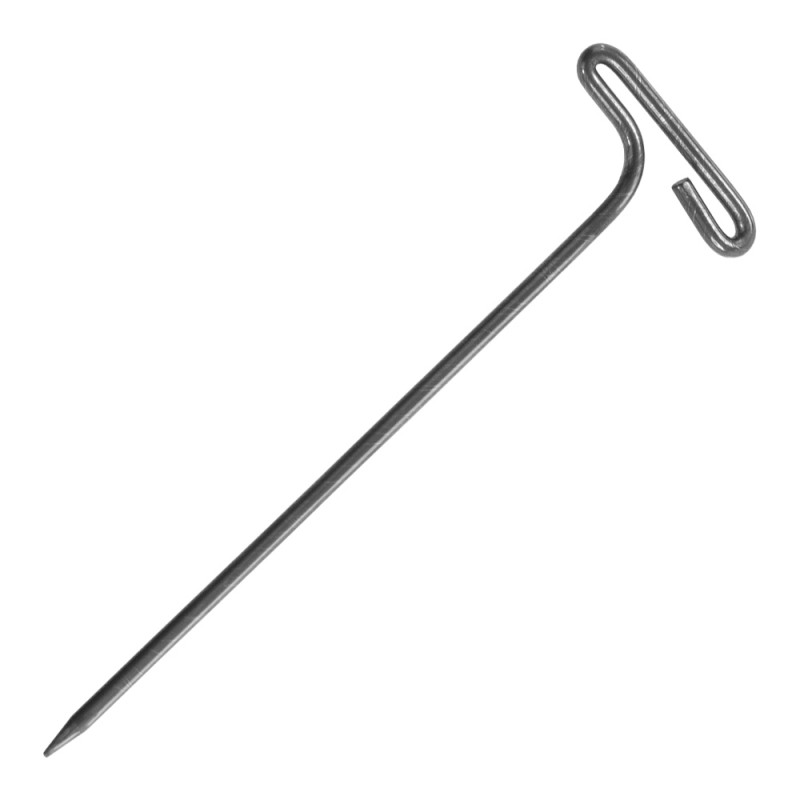
Left): Porcine ventricle sample, epicardium side up, mounted to
Download scientific diagram | (Left): Porcine ventricle sample, epicardium side up, mounted to the silicone lined fixture with Tpins. (Right): Porcine aorta sample, intima side up, mounted to the silicone lined fixture with T-pins. (Both): 0.25 in diameter steel ball upper member as test probe. from publication: PolyJet 3D Printing of Tissue Mimicking Materials: An Investigation of Characteristic Properties of 3D Printed Synthetic Tissue | Current anatomical 3D printing has been primarily used for education, training, and surgical planning purposes. This is largely due to the models being printed in materials which excel at replicating macro-level organic geometries; however, these materials have the drawback | 3D Printing, Tissue and Subcutaneous Tissue | ResearchGate, the professional network for scientists.

Spatial and temporal patterns of SAN and atrial genes indicate
Frontiers Porcine Organotypic Epicardial Slice Protocol: A Tool

Emily A. Bermel's research works University of Minnesota Duluth

Section levels of the left ventricle.

Intramural Needle Ablation for Refractory Premature Ventricular

PDF) PolyJet 3D Printing of Tissue Mimicking Materials: An

Physiological Biomimetic Culture System for Pig and Human Heart

Heart Anatomy Anatomy and Physiology II

Epicardial slices: an innovative 3D organotypic model to study

PDF) PolyJet 3D Printing of Tissue Mimicking Materials: An
Bioengineering, Free Full-Text

Wide-area low-energy surface stimulation of large mammalian

The Left and Right Ventricles